Aging and age-related diseases profoundly affect stem cell proliferation and differentiation, which has implications for tissue homeostasis. The bone marrow niche maintains and regulates stem cells and interacts with endothelial cell subtypes. It plays a key role in mediating bone homeostasis by recruiting bone cells. During ageing and under pathological conditions, detrimental changes occur in the bone marrow niche that affect the microvascular system and lower blood flow. This is exacerbated in type 2 diabetes, a disease characterised by hyperglycaemia and high levels of inflammation.
This project brings together the expertise of the University Hospital Hamburg-Eppendorf (UKE), the University of Southern Denmark (SDU) and the German Electron Synchrotron facility (DESY). The collaboration combines expertise in bone tissue quality analysis, bone histology and high-resolution imaging, and is located close to the clinics. The project will use advanced imaging and state-of-the-art tissue analysis techniques to determine changes in the vasculature of the bone marrow in patients with type 2 diabetes. The research team will conduct 3D nano-tomographic imaging of vertebral samples from individuals with type 2 diabetes and controls to visualise the microvascular network within the human bone marrow niche. Bone histology will be used to characterise vessel type and bone marrow stem cells. This study will provide new technological approaches and insights into the underlying mechanisms of diabetes and vascular changes.
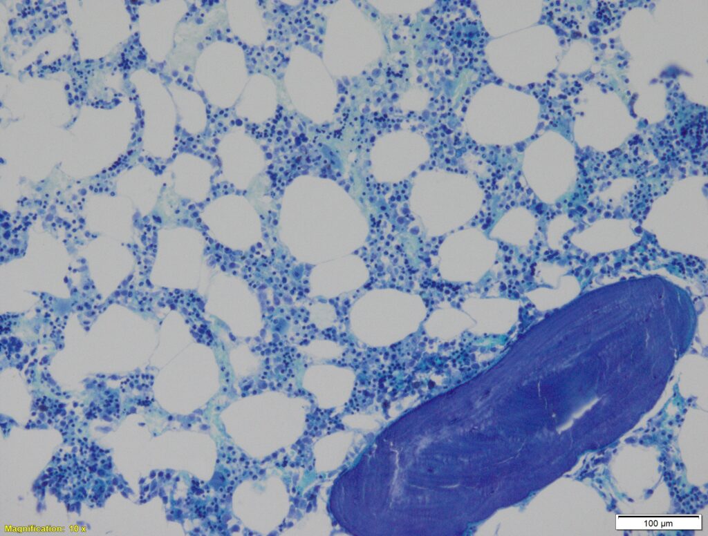
The figure shows adipocyte ghosts within the intertrabecular spaces. Bone marrow sinusoids are lined by endothelial cells resting on a discontinuous basement membrane. In the bone marrow, sinusoids are often collapsed and therefore challenging to visualise.
